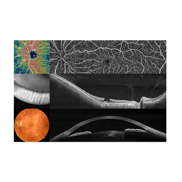
SOLIX FullRange anterior imaging provides stunning views of the entire anterior chamber, from the front surface of the cornea to the anterior surface of the lens. A comprehensive anterior segment package expands the clinical utility of the system to address a broad range of patients.
Images of the entire anterior chamber can be captured with the FullRange 18 x 6.25mm scan and ocular structures can be measured with the caliper tools.
Corneal Layer Map
Corneal & epithelial mapping
Epithelial, stromal and total corneal thickness can be quantified with the 10mm Corneal Layer Map, which features 16 meridians to fully cover the LRS transition zone. The Highlight Tool shows any subtle changes in thickness and the Change Analysis report measures changes in thickness between visits.
Epithelial, stromal and total corneal thickness can be measured to aid in disease diagnosis. Pachymetric measurements may be compared to the Coollabs Keratoconus Risk Scoring System to further enhance diagnostic accuracy.
External IR imaging
Evaluate Meibomian glands
SOLIX's infrared capabilities allow you to evaluate Meibomian gland structure of the upper and lower lids, which means new information can be added to the diagnosis and management of dry eye patients, without the need for a dedicated imaging system.
FullRange retina
Wide & deep imaging
SOLIX delivers pristine images of retinal structures with the 16mm x 6.25mm FullRange retina scan that enables confident diagnosis and management of retinal pathologies, even in highly myopic patients. Diagnostic capabilities can be enhanced further with an ultra-fast, deep and wide line scan.
FullRange retina – dual montage
En Face OCT
A single scan protocol with MCT generates all necessary images and data for comprehensive retinal analysis, which optimises efficiency and quickly provides the clinical data your practice demands. Two 9 x 9mm scans can be combined for a seamless view of the posterior pole.
Photography
Fundus & external
The fundus photography application enables the capture of a 45-degree fundus image, as well as the ability to view retinal photos in color, grayscale and inverse modes, with a range of tools, including a cup-to-disc ratio measuring tool.
Glaucoma analytics
Combine structural & vascular images
The SOLIX glaucoma package delivers in-depth analysis of the optic nerve head structure and vasculature. Optovue-exclusive data points bring additional insights that aid in clinical decision making.
Glaucoma - trend analysis
Assess change
ONH + GCC Trend Analysis Report
Track change and estimate the rate of change in both GCC and RNFL thickness with unparalleled reproducibility to easily assess how quickly a patient’s disease is progressing.
AngioDisc Trend Analysis Report
Measure the vessel density of the RPCs, assess visit-to-visit change and estimate rate of change in glaucoma patients and suspects. Vessel density analysis complements RNFL and GCC analysis and aids in the management of advanced glaucoma – especially in cases where neural structural measurements have reached the measurement floor.