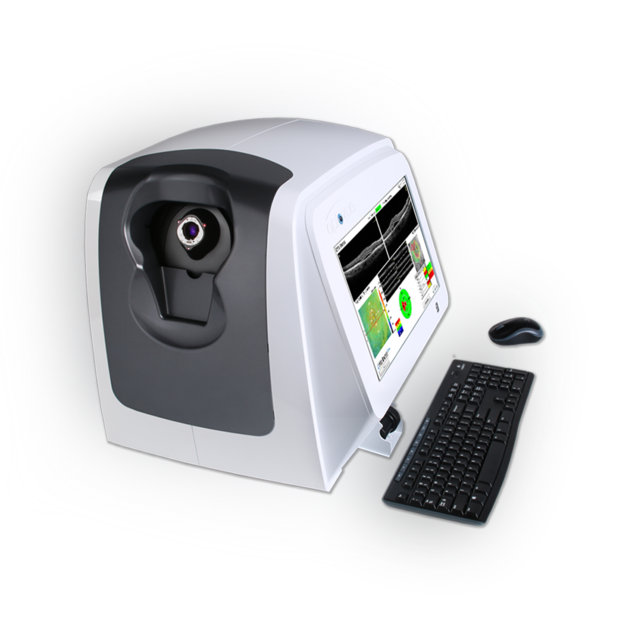
Fully-integrated OCT system

Comprehensive suite of scans

Retinal imaging

Full range of retina scans

Real-time en face display provides a 12x9mm view of the retina during scan acquisition to assist the operator in scanning the desired location. 3D 7x7mm retina cube scan provides visualization of 201 raster lines to enable in-depth analysis of retinal structures. Six 12mm radial lines provide multiple views of the retina. Retina Map allows you to visualize a 9x5mm area of the retina, with an ETDRS reference database comparison, to quickly identify areas of increased or decreased thickness.

Optic nerve

Extensive glaucoma scans

A 6x6mm 3D disc cube scan provides visualization of 201 raster lines to enable in-depth analysis of optic disc structures. The Ganglion Cell Complex (GCC) thickness map allows identification and measurement of ganglion cell loss in glaucoma and Optovue’s exclusive Focal Loss Volume metric (FLV%) is the single best predictor of conversion to glaucoma. The nerve fiber thickness map allows visualization and quantification of RNFL thinning in glaucoma. iScan80’s GCC and RNFL analysis reports include single eye and OU reports, change analysis for visit-to-visit comparison, trend analysis to assess change over time and combo reports that display both GCC and RNFL thickness profiles for comprehensive analysis.

Anterior segment

Comprehensive range of anterior scans

Visualize and quantify 6mm of epithelial, stromal and total corneal thickness to identify areas of thickening or thinning related to dry eye disease, keratoconus, or previous refractive surgery. The Change Analysis report measures changes in thickness between visits. Vault Mapping allows you to visualize the fluid reservoir between the lens and cornea for precise scleral lens fitting. Assess angle structure with a quick, non-contact scan and quantify angle parameters with easy-to-use measurement tools.
Epithelial Thickness Mapping (ETM)

In-depth analysis

The anterior segment module offers a 6mm diameter epithelial thickness map with minimum thickness marker. Epithelial Thickness Mapping (ETM) aids the clinician in evaluating potential Keratoconus, LASIK/LASEK/PRK planning, tracking epithelial regrowth, dry eye and contact lens patients.

Further capabilities

Specialty contact lens fitting

The Vault Mapping tool enables precise visualization of the lens-to-eye relationship for specialty lens fitting. The user can evaluate central corneal clearance in exact microns with the vault map report, and measure lens settling between the first and second scan with the change map. Superior, temporal, inferior and nasal landing zones can also be evaluated with the anterior display report.