Biometry
Anterior Chamber Suite
Comprehensive data & visualization for corneal diagnostics


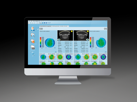
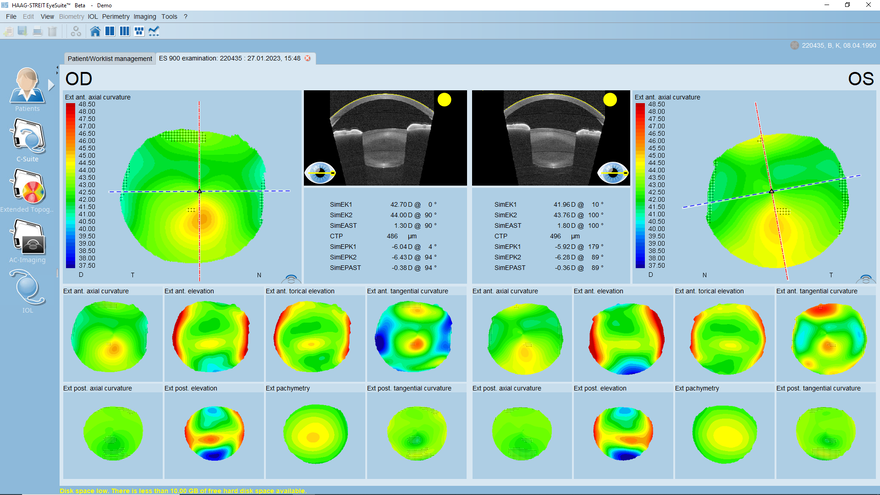
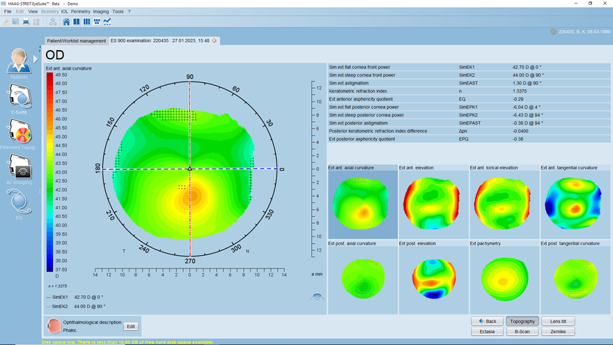
Overview screen
Comprehensive data for thorough diagnosis and clear visualization
Thanks to its cutting-edge swept source OCT technology, the Anterior Chamber Suite provides class A-topography of up to 12 mm diameter of the anterior and posterior corneal surface, and OCT imaging of the anterior segment, including the lens and the chamber angle, with up to 18 mm diameter coverage in a comprehensive overview screen
Topography detail view
Review and follow up in detail
The topography detail screen of each measurement is accessed by clicking on any of the measurements or displays on the overview screen. It allows a more thorough and detailed review of the respective information in separate displays providing more information and additional analysis tools.
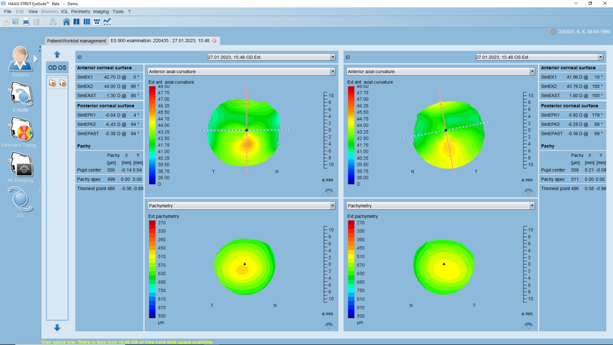
Four-in-one view
Comparison of maps and measurements
The four-in-one view enables the user to review two maps and the corresponding measurement data from either two points in time or between OD and OS. The maps reviewed can be fully defined by the user and may be changed with a simple click while looking at the data.
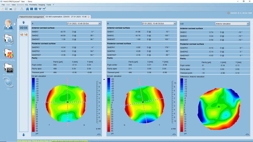
Table difference view
Quantify differences
In the table difference view two measurements A and B (two displays on the left) from either two points in time or OD/DS data from the same point in time are displayed as a difference view showing the difference between A and B. The differences are displayed both quantitatively as well as in a single, intuitive visualization, that can be individually adjusted.
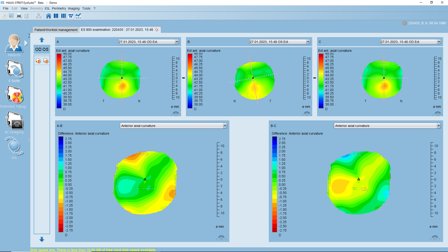
Map difference view
Visualization of differences
In the map difference view the user may review changes in up to three data points in any of the map types available on the Eyestar 900. On the top, the three data points A, B and C are displayed, while at the bottom the difference between A and B as well as the difference between B and C are shown. This enables identification of changes in an intuitive way.
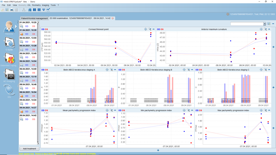
Progression view
Visualization of progression
The progression view enables the user to create its own progression display for any data point available on the Eyestar. The user might create reports for specific diagnostic means, incorporating the parameters the user is most interested in.
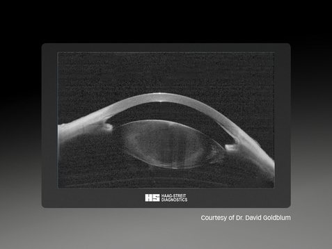
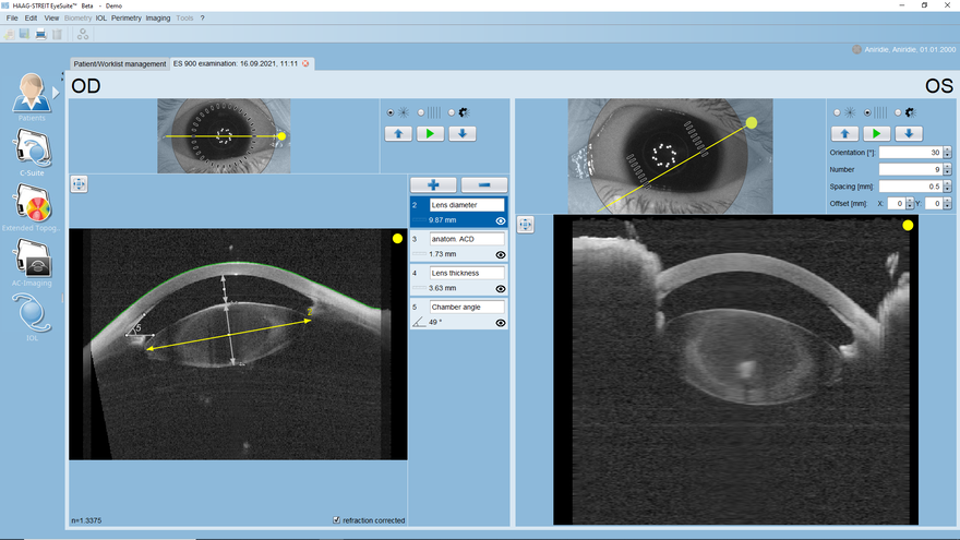
18 mm Swept-Source OCT Imaging
Revealing hidden details
The Anterior Chamber Suite imaging mode provides OCT imaging of the anterior segment, including the lens and the chamber angle, with up to 18 mm diameter coverage. Due to image acquisition using the cutting-edge, patent-protected Mandala scan technology, virtual B-scans may be created anytime and anywhere in the already acquired volume, minimizing the need for the rescanning of patients to visualize details. Manual measurements tools for distances, angles and areas complement this unique imaging and documentation feature of Eyestar 900.

Overview screen

Topography detail view

Four-in-one view

Table difference view

Map difference view

Progression view

18 mm Swept-Source OCT Imaging


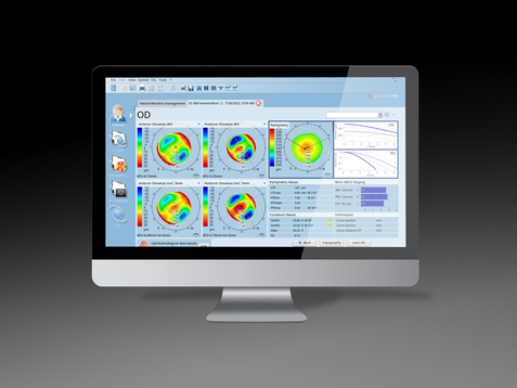
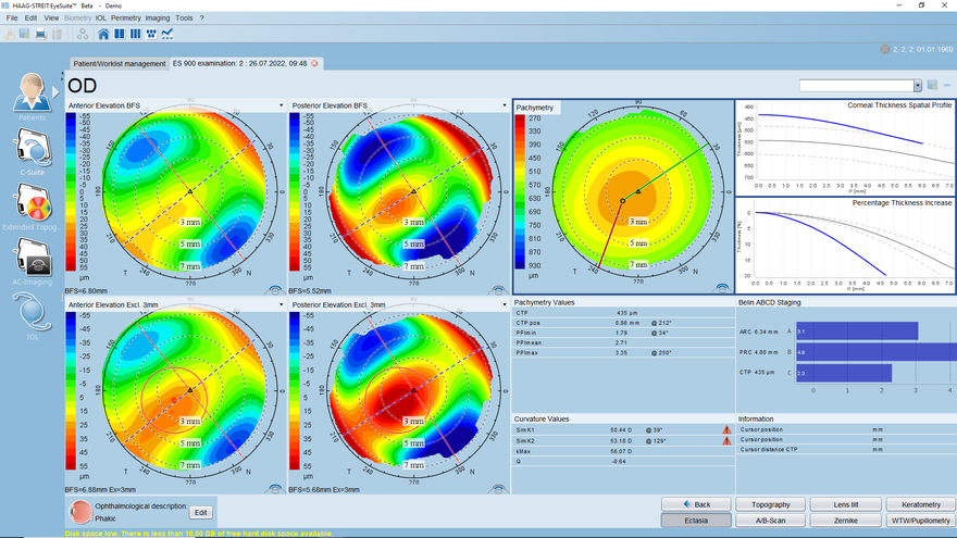
Corneal Ectasia Display
Excellent tool to detect corneal ectasia
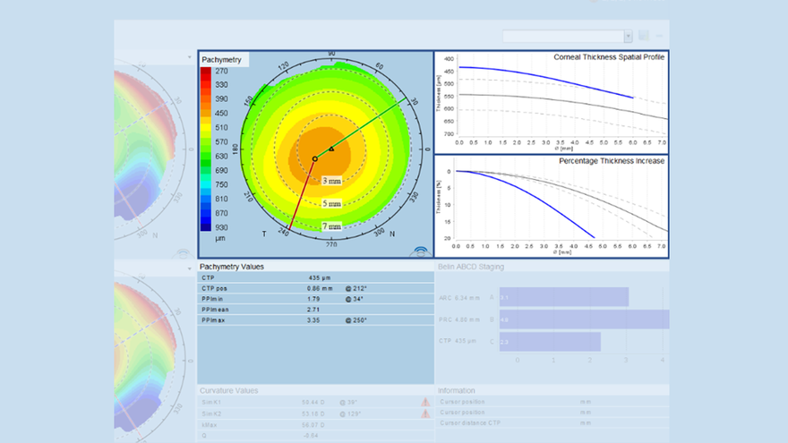
The corneal ectasia display provides a comprehensive and customizable view for identification of early signs of corneal ectatic changes. On the left, four displays including power, elevation and pachymetry maps can be individually chosen. It further offers a pachymetry map and progression graphs on the top right as well as the Belin ABCD grading underneath. The corneal ectasia display is further complemented with additional parameters like simK and Kmax, providing a wealth of information for thorough diagnosis.
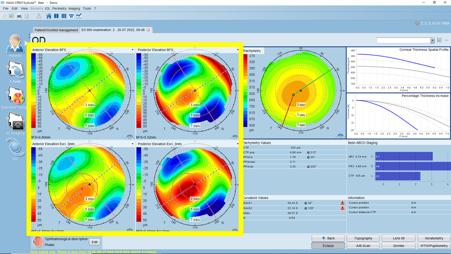
Elevation map for enhanced visualization
Intuitive identification of ectatic change
In addition to power, elevation and pachymetry maps, there is also an elevation map available on which the best fit sphere is calculated excluding an area of 3mm diameter around the thinnest point of the cornea. This exclusion of a potential pathologically affected area may improve the visualisation of such an ectatic change in the map.
Pachymetry map & progression
Identify unusual corneal pachymetry progression
Unusual quick thickening of the cornea may be a sign of developing keratoconus. For easier identification, a pachymetry map and two progression graphs are shown. The progression graphs include a normative reference graph of a healthy Caucasian population with a 95% confidence interval to identify unusual progression of corneal thickness. The data is further complemented with the average, the maximum and minimum Pachymetry Progression Indices. The more these numbers differ form 1 (normal) the higher the likelihood of corneal pathology.
Belin ABCD grading
Gold-standard keratoconus grading system
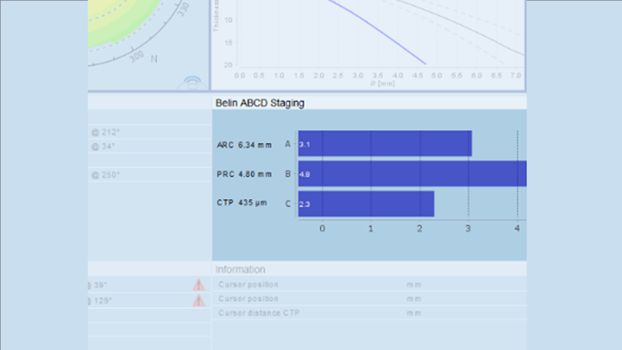
The Belin ABCD Keratoconus grading system is the gold standard in staging the severity of keratoconus in a patient. With its intuitive four parameter-based scoring, you can confidently assess the severity of your patients' condition and provide them with the best possible treatment options. The four parameters are A for the anterior corneal radius (ARC), B for the posterior cornea radius (PRC), C for the thinnest point of the cornea (CPT) and D for the distance visual acuity (VA). Its performance has been documented in several peer-reviewed articles.

Corneal Ectasia Display

Elevation map for enhanced visualization

Pachymetry map & progression

Belin ABCD grading
